AE-Adult-Echocardiography Valid Exam Braindumps | AE-Adult-Echocardiography Test Simulator Free
Wiki Article
What's more, part of that SurePassExams AE-Adult-Echocardiography dumps now are free: https://drive.google.com/open?id=1D5PcnuP19e0prv2PzAWIW6xK78F2lMcJ
SurePassExams is famous for high-quality reliable exam bootcamp materials recent years. Our valued customers enjoy the privilege: pass guaranteed; our AE-Adult-Echocardiography study guide materials find the best meaning in those candidates who have struggled hard to pass the AE-Adult-Echocardiography certification exams. We have special information resources about many international companies. We promise most Reliable AE-Adult-Echocardiography Exam Bootcamp materials are the latest version which are edited based on first-hand information. You can rest assured to purchase our AE-Adult-Echocardiography study guide materials.
ARDMS AE-Adult-Echocardiography Exam Syllabus Topics:
| Topic | Details |
|---|---|
| Topic 1 |
|
| Topic 2 |
|
| Topic 3 |
|
| Topic 4 |
|
| Topic 5 |
|
>> AE-Adult-Echocardiography Valid Exam Braindumps <<
AE-Adult-Echocardiography Test Simulator Free & AE-Adult-Echocardiography Learning Engine
The AE Adult Echocardiography Examination certification exam is one of the top-rated career advancement certification exams. The ARDMS AE-Adult-Echocardiography certification exam can play a significant role in career success. With the AE Adult Echocardiography Examination (AE-Adult-Echocardiography) certification, you can gain several benefits such as validation of skills, career advancement, competitive advantage, continuing education, and global recognition of your skills and knowledge.
ARDMS AE Adult Echocardiography Examination Sample Questions (Q110-Q115):
NEW QUESTION # 110
Which kind of cardiac valve is a heterograft?
- A. One that is from one location to another in the same human
- B. One that is from pericardial tissue
- C. One that is from an animal to a human
- D. One that is from a human to another human
Answer: C
Explanation:
A heterograft (also called xenograft) cardiac valve is derived from an animal species, commonly porcine or bovine, and implanted into a human. These bioprosthetic valves are treated to reduce immunogenicity.
Option A describes an allograft (homograft). Option B refers to bioprosthetic valves but does not specify species. Option C describes an autograft, such as the Ross procedure.
This classification is standard in cardiac surgery and echocardiography literature#16:Textbook of Clinical Echocardiography, 6ep.450-455##12:ASE Valve Prosthesis Guidelinesp.200-205#.
NEW QUESTION # 111
Which of the following are key features of an unrepaired tetralogy of Fallot?
- A. Displaced tricuspid valve, atrialization of the right ventricle, severe tricuspid regurgitation, and a secundum atrial septal defect
- B. Supravalvular mitral valvular ring, subaortic membrane, bicuspid aortic valve, and aortic coarctation
- C. Outlet ventricular septal defect, overriding aorta, right ventricular outflow tract obstruction, and right ventricular hypertrophy
- D. Inlet ventricular septal defect, common atrioventricular valve, atrioventricular valve regurgitation, and primum atrial septal defect
Answer: C
Explanation:
Comprehensive and Detailed Explanation From Exact Extract:
Tetralogy of Fallot (TOF) is a congenital heart defect characterized by four key anatomical abnormalities: an outlet (malalignment) ventricular septal defect (VSD), an overriding aorta that receives blood from both ventricles, right ventricular outflow tract (RVOT) obstruction (commonly infundibular stenosis), and resultant right ventricular hypertrophy. These defects cause cyanosis due to right-to-left shunting and impaired pulmonary blood flow.
Option A describes Ebstein anomaly, characterized by a displaced tricuspid valve and atrialization of the right ventricle.
Option B describes features more consistent with Shone complex or other left heart obstructive lesions.
Option C describes atrioventricular septal defect (AVSD), seen in conditions like Down syndrome.
In unrepaired TOF, echocardiography demonstrates the large malalignment VSD, overriding aorta, RVOT obstruction, and hypertrophied right ventricle. These are classic textbook findings described in adult and pediatric echocardiography literature, including "Textbook of Clinical Echocardiography" (Chapter on Congenital Heart Disease) and ASE guidelines#16:Textbook of Clinical Echocardiography, 6ep.560-565#
#12:ASE Adult Congenital Guidelinesp.400-410#.
NEW QUESTION # 112
Which adjustment is most likely to improve image quality from the suprasternal long axis window?
- A. Ask patient to look slightly toward the left
- B. Rotate transducer indicator toward the patient's right shoulder
- C. Move probe just inferior to the sternum
- D. Place patient in left lateral decubitus position
Answer: A
Explanation:
Comprehensive and Detailed Explanation From Exact Extract:
The suprasternal long axis window is best accessed with the patient in the supine position with the neck extended. To optimize image quality, instructing the patient to turn their head slightly toward the left side moves the trachea and clavicle away from the ultrasound beam path, allowing better visualization of the aortic arch and great vessels.
Moving the probe inferior to the sternum accesses the subxiphoid window rather than suprasternal. Left lateral decubitus improves parasternal and apical windows but not suprasternal. Rotating the transducer indicator toward the patient's right shoulder would change the imaging plane but is not a primary method to improve image quality.
This technique is highlighted in the "Textbook of Clinical Echocardiography, 6e", Chapter on Echocardiographic Windows and Acoustic Access#20:90-95Textbook of Clinical Echocardiography#.
NEW QUESTION # 113
Which condition is most plausible based on the finding indicated by the arrow on this image?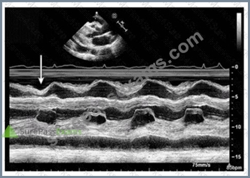
- A. Constrictive pericarditis
- B. Cardiac tamponade
- C. Pulmonary hypertension
- D. Pulmonary embolism
Answer: A
Explanation:
The image is a parasternal long axis M-mode echocardiographic tracing demonstrating the interventricular septum and posterior left ventricular wall. The arrow points to the septal "bounce" or "shudder," which is an abnormal early diastolic septal motion.
This septal bounce is a classic echocardiographic finding in constrictive pericarditis, caused by rapid early diastolic filling with abrupt cessation due to pericardial constraint, resulting in paradoxical septal motion.
Cardiac tamponade usually shows pericardial effusion with chamber collapse but not septal bounce.
Pulmonary embolism and pulmonary hypertension have different echocardiographic signs such as right ventricular dilatation and pressure overload but no septal bounce.
These features are well described in the "Textbook of Clinical Echocardiography" and ASE pericardial disease guidelines#16:Textbook of Clinical Echocardiography, 6ep.280-285##12:ASE Pericardial Disease Guidelinesp.300-305#.
NEW QUESTION # 114
A patient with a ventricular septal defect, an atrial septal defect, and a cleft mitral valve is likely to have which abnormality?
- A. Ebstein anomaly
- B. Atrioventricular canal defect
- C. Shone syndrome
- D. Marfan syndrome
Answer: B
Explanation:
Comprehensive and Detailed Explanation From Exact Extract:
Atrioventricular canal defect (AV canal defect) is a congenital cardiac malformation characterized by defects in the atrial and ventricular septa, along with abnormalities of the atrioventricular valves including cleft mitral valve. These features collectively cause shunting and valve regurgitation.
Ebstein anomaly primarily involves the tricuspid valve and right atrium, Marfan syndrome is a connective tissue disorder with different manifestations, and Shone syndrome involves left-sided obstructive lesions.
This is clearly outlined in the "Textbook of Clinical Echocardiography, 6e", Chapter on Congenital Heart Defects - Atrioventricular Septal Defects#20:120-125Textbook of Clinical Echocardiography#.
NEW QUESTION # 115
......
The SurePassExams is a leading platform that is committed to offering to make the ARDMS Exam Questions preparation simple, smart, and successful. To achieve this objective SurePassExams has got the services of experienced and qualified AE Adult Echocardiography Examination (AE-Adult-Echocardiography) exam trainers. They work together and put all their efforts and ensure the top standard of SurePassExams AE Adult Echocardiography Examination (AE-Adult-Echocardiography) exam dumps all the time.
AE-Adult-Echocardiography Test Simulator Free: https://www.surepassexams.com/AE-Adult-Echocardiography-exam-bootcamp.html
- Reliable AE-Adult-Echocardiography Test Prep ???? New AE-Adult-Echocardiography Test Braindumps ???? AE-Adult-Echocardiography Test Prep ⬅️ Enter ✔ www.validtorrent.com ️✔️ and search for 「 AE-Adult-Echocardiography 」 to download for free ????AE-Adult-Echocardiography Certification Test Answers
- AE-Adult-Echocardiography Latest Torrent ???? AE-Adult-Echocardiography Certification Test Answers ???? AE-Adult-Echocardiography Test Registration ❓ Easily obtain free download of ➥ AE-Adult-Echocardiography ???? by searching on 【 www.pdfvce.com 】 ????New AE-Adult-Echocardiography Test Forum
- AE-Adult-Echocardiography Valid Exam Braindumps - 100% Pass Quiz 2026 AE-Adult-Echocardiography: AE Adult Echocardiography Examination First-grade Test Simulator Free ???? Copy URL 「 www.troytecdumps.com 」 open and search for ▛ AE-Adult-Echocardiography ▟ to download for free ????AE-Adult-Echocardiography Answers Real Questions
- AE-Adult-Echocardiography Exam Lab Questions ???? Certification AE-Adult-Echocardiography Test Answers ???? AE-Adult-Echocardiography Answers Real Questions ???? Easily obtain free download of 【 AE-Adult-Echocardiography 】 by searching on 《 www.pdfvce.com 》 ⭐AE-Adult-Echocardiography Test Prep
- Free PDF ARDMS - AE-Adult-Echocardiography - High Pass-Rate AE Adult Echocardiography Examination Valid Exam Braindumps ???? Immediately open ➡ www.dumpsquestion.com ️⬅️ and search for ▛ AE-Adult-Echocardiography ▟ to obtain a free download ????AE-Adult-Echocardiography Real Dump
- ARDMS AE-Adult-Echocardiography Valid Exam Braindumps: AE Adult Echocardiography Examination - Pdfvce Latest updated ???? Search for ➤ AE-Adult-Echocardiography ⮘ and download it for free immediately on ( www.pdfvce.com ) ????AE-Adult-Echocardiography Latest Torrent
- New AE-Adult-Echocardiography Test Forum ???? AE-Adult-Echocardiography Latest Braindumps Sheet ???? AE-Adult-Echocardiography Exam Actual Questions ☃ Go to website “ www.testkingpass.com ” open and search for ▛ AE-Adult-Echocardiography ▟ to download for free ????AE-Adult-Echocardiography Certification Test Answers
- AE-Adult-Echocardiography Exam Actual Questions ???? AE-Adult-Echocardiography Real Dump ???? Download AE-Adult-Echocardiography Free Dumps ???? Download [ AE-Adult-Echocardiography ] for free by simply searching on ☀ www.pdfvce.com ️☀️ ????AE-Adult-Echocardiography Real Dump
- Exam AE-Adult-Echocardiography Success ???? New AE-Adult-Echocardiography Test Braindumps ???? AE-Adult-Echocardiography Real Dump ???? Open ☀ www.verifieddumps.com ️☀️ enter ✔ AE-Adult-Echocardiography ️✔️ and obtain a free download ????Download AE-Adult-Echocardiography Free Dumps
- New AE-Adult-Echocardiography Test Topics ???? Certification AE-Adult-Echocardiography Test Answers ???? AE-Adult-Echocardiography Certification Test Answers ???? Open [ www.pdfvce.com ] enter 【 AE-Adult-Echocardiography 】 and obtain a free download ????Download AE-Adult-Echocardiography Free Dumps
- New AE-Adult-Echocardiography Test Forum ???? AE-Adult-Echocardiography Answers Real Questions ???? New AE-Adult-Echocardiography Test Pattern ???? Search for ⏩ AE-Adult-Echocardiography ⏪ and obtain a free download on ▛ www.dumpsmaterials.com ▟ ????AE-Adult-Echocardiography Test Prep
- lewistsfo672058.bloguerosa.com, myportal.utt.edu.tt, myportal.utt.edu.tt, myportal.utt.edu.tt, myportal.utt.edu.tt, myportal.utt.edu.tt, myportal.utt.edu.tt, myportal.utt.edu.tt, myportal.utt.edu.tt, myportal.utt.edu.tt, myportal.utt.edu.tt, www.stes.tyc.edu.tw, alexisimport.com, laytnjumk368567.blogoxo.com, socialaffluent.com, myportal.utt.edu.tt, myportal.utt.edu.tt, myportal.utt.edu.tt, myportal.utt.edu.tt, myportal.utt.edu.tt, myportal.utt.edu.tt, myportal.utt.edu.tt, myportal.utt.edu.tt, myportal.utt.edu.tt, kingbookmark.com, haarisggxd599970.blogcudinti.com, violagaeh140815.thenerdsblog.com, Disposable vapes
What's more, part of that SurePassExams AE-Adult-Echocardiography dumps now are free: https://drive.google.com/open?id=1D5PcnuP19e0prv2PzAWIW6xK78F2lMcJ
Report this wiki page